
The Importance of Biopsy after Lump Removal: Ensuring Clear Margins and Cancer-Free Outcomes

Removing a lump from your pet is a critical step, but it is only part of the process. Many pet owners assume that once the lump is gone, the problem is solved. The truth is, without a biopsy, we cannot be sure if the surgery was successful in removing all the abnormal tissue. This uncertainty can affect your pet’s health and future well-being.
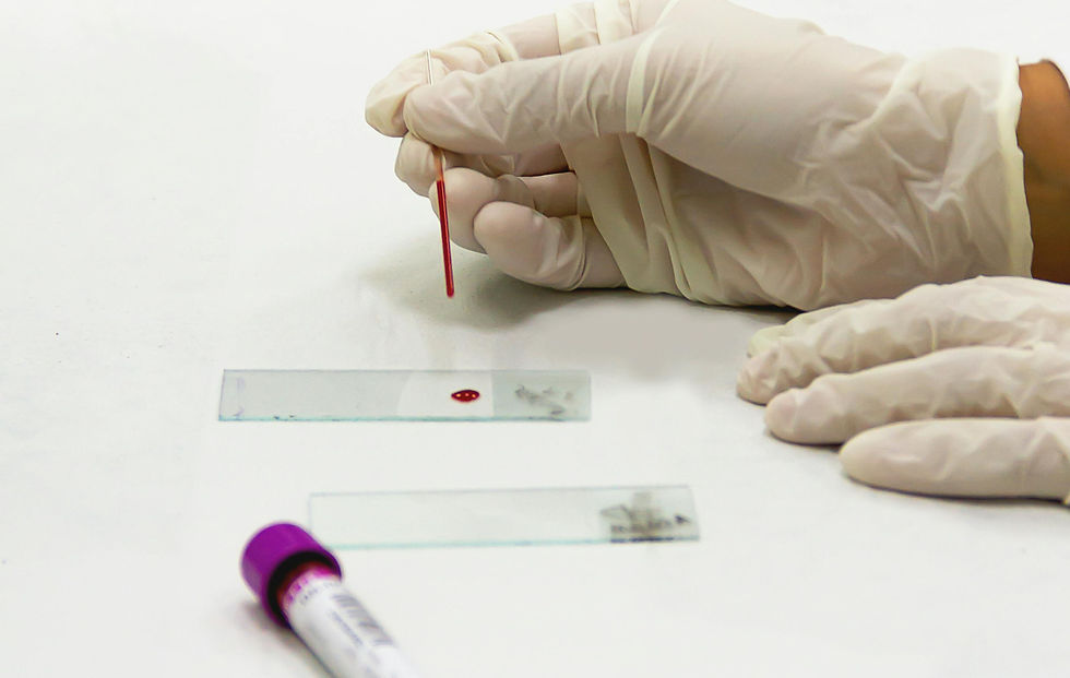
Why Removing the Lump Is Only Half the Job
When a lump is surgically removed, the visible mass is taken out, but cancer cells can sometimes remain at the edges of the removed tissue. These leftover cells can cause the cancer to come back. A biopsy, also called histology, examines the lump under a microscope to check if the edges, or margins, are “clean.” Clean margins mean no cancer cells are found at the edges, suggesting the entire lump was removed.
Without this detailed examination, we cannot know if the surgery was complete.
What Does a Biopsy Involve?
After the lump is removed, the tissue is sent to a laboratory where a pathologist examines it. The pathologist looks for:
The type of cells in the lump
Whether the lump is benign (non-cancerous) or malignant (cancerous)
If cancer cells are present at the edges of the tissue
This information helps the veterinarian plan the next steps. For example, if cancer cells are found at the margin, your pet may need another surgery or additional treatments like chemotherapy.
What Pet Owners Should Know
Always ask for a biopsy after lump removal.
Understand that biopsy results take time, usually a few days to a week.
Follow your veterinarian’s advice based on the biopsy report.
Keep an eye on the surgery site for any changes or new lumps.


Comments